
New fMRI evidence for hyperfocusing in schizophrenia
/Hahn, B., Bae, G.-Y., Robinson, B. M., Leonard, C. J., Luck, S. J., & Gold, J. M. (2020). Cortical hyperactivation at low working memory load: A primary processing abnormality in people with schizophrenia? NeuroImage: Clinical, 26, 102270. https://doi.org/10.1016/j.nicl.2020.102270
The hyperfocusing hypothesis proposes that schizophrenia involves an abnormally narrow but intense focusing of processing resources. That is, people with schizophrenia are not impaired at focusing their attention; on the contrary, they tend to focus their attention more intensely and more narrowly compared to healthy control subjects. I wrote a blog post about this hypothesis last September (when we published a couple review papers). Today, I’d like to describe evidence from a new fMRI study of visual working memory—led by Britta Hahn—that was recently published in NeuroImage: Clinical (open access!).
The bottom line: When we compared groups of people with schizophrenia (PSZ) and healthy control subjects (HCS) who were matched for behavioral performance, PSZ exhibited greater BOLD activation than HCS when required to maintain a single object in working memory. This is exactly what we mean by a “more intense” focusing of processing resources in PSZ.
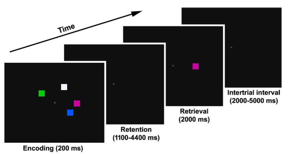
Now for the details. As shown in the diagram, subjects performed a change detection task. On each trial, subjects were shown an Encoding Array consisting of 1–7 colored squares, which they were required to maintain in working memory over a delay period. Then, a colored square was shown at one of the original locations, and subjects had to indicate whether this was the same color as the square that had been present at that location in the Encoding Array. Tons of previous research has shown that this task is an excellent measure of working memory capacity (reviewed here).
Previous fMRI research by Todd & Marois (2004, 2005) has shown that the posterior parietal cortex (PPC) plays a key role in this task: BOLD activity in PPC increases as the number of items maintained in WM increases. Because this task does not involve significant manipulation or interference, the PFC does not appear to play much role. Just like Todd & Marois, we found an area of the PPC in which the BOLD signal varied across set size in a manner that matched the number of objects that were stored in memory (as measured behaviorally). This result was previously published, along with evidence that PSZ exhibited greater BOLD activity than HCS at set size 1 (consistent with hyperfocusing). This is consistent with a previous ERP study led by Carly Leonard, in which we found greater delay-period activity in PSZ than in HCS at set size 1.
However, it can be problematic to compare fMRI data across patients and controls when they also differ in performance. For example, if PSZ have decreased working memory capacity, then this may cause them to exert more effort at low set sizes, so the greater BOLD activity at set size 1 in PSZ relative to HCS could be a side effect of lower working memory capacity in PSZ. In our previous ERP study, we addressed this by comparing subsets of PSZ and HCS who were matched on behaviorally measured working memory capacity (K). Our new fMRI paper took this same approach. We ended up with subgroups of 23 PSZ and 23 HCS (from an original sample of 37 PSZ and 37 HCS) that were very well matched on behavioral performance.

The results are summarized in the figure. You can see the area of posterior parietal cortex that was related to working memory capacity, the BOLD signal at each set size in the full groups (A), and the BOLD signal at each set size in the matched subgroups (B). Whether or not we matched performance, the BOLD signal was much larger in PSZ than in HCS at set size 1, consistent with “an abnormally narrow but intense focusing of processing resources” in schizophrenia.